Research Terms
Energy Consumption Energy Technology Nuclear Energy Fission Energy Fusion Energy Nuclear Fuels Materials Sciences Nuclear Engineering Nuclear Accelerators Nuclear Power Plants Nuclear Materials Transport Spent Fuel Storage Nuclear Safety
Industries
Goal: Creating nuclear safeguard-enabling detectors, neutron interaction understandings, and ground-breaking novel data approaches. Application: Using data fusion between 3D-computer vision and radiation sensors to detect and track potential threats as imposed by lost or smuggled radiation sources.
Subject Areas:
Audience:
Adults
Duration:
1 hour or less
Fee:
Less than $500
Goal: Creating nuclear safeguard-enabling detectors, neutron interaction understandings, and ground-breaking novel data approaches. Application: Using data fusion between 3D-computer vision and radiation sensors to detect and track potential threats as imposed by lost or smuggled radiation sources.
Subject Areas:
Audience:
High School
Duration:
1 hour or less
Fee:
Expenses Only

| Year: | 2015 |
| Link Address: | MHCQz7ySgKo |
| Keywords: | safeguards, detection, radiation, neutron |
| Source: | youtube |
| Duration: | 00:15:00 |
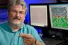
This radiological threat detection system uses 3D-vision tracking data to improve accuracy and allow easier detection of materials with lower radiation levels. Radiological detection systems can enable nuclear facility operations, help secure high traffic public places, or recover lost or stolen nuclear material. Common security systems such as radiation portal monitors or sensor networks that detect potential radiological threats are also prone to false alarms that waste time, money, and resources. In addition, current systems face challenges when trying to maintain a continuity of commerce and people in busy, dynamic environments. On the other hand, avoiding alarms due to background radiation or non-threatening sources requires setting high detection thresholds, which may miss potential threats with weaker radiation signals.
Researchers at the University of Florida have developed a radiation monitoring system solution that has increased accuracy and better sensitivity to detect weaker sources and reduce the false positive rate. Integrating spatial and visual data in determining potential threats also allows a system to track radiation sources in high traffic areas.
High-accuracy radiation scanning system that detects and tracks radiological material to enhance security at airports, seaports, shipping facilities, train stations, government buildings, etc. Active algorithm and sensor upgrade option for existing radiation monitoring solutions.
This detection system uses an algorithm to fuse data from 3D-vision sensors and radiological detectors in order to improve the accuracy of identifying potentially threatening radiation sources. Visual tracking provides contextual data from the scene that informs the radiation scanning process, causing fewer false positives. The better accuracy allows reliable detection using a lower threshold, allowing security systems to be more sensitive to lower radiation levels. The system solution allows operation and sensitivities significantly below the standard Currie-limit of standard previous detection systems.
This radiographic monitoring system tracks particle beam delivery within a patient undergoing particle therapy. The goal of radiation therapy is to maximize the treatment dosage delivered to a tumor while minimizing exposure of surrounding healthy tissue. Studies have shown that charged particle therapies such as proton therapy and heavy ion therapy significantly reduce toxicity to normal tissue while focusing radiation on a tumor. However, real-time guidance in charged particle therapy is limited by the finite range of the charged particles, which makes it difficult to determine the precise position of the tumor target within the beam aperture.
Higher energy particles during the interaction with matter can produce secondary particles such as neutrons, electrons, x-rays and gammas. Neutrons and photons, which tend to be more penetrating than the charged particles, typically give the largest contribution to the secondary dose in out-of-field organs. Researchers at the University of Florida have developed a portal imaging system for charged particle therapy based on real-time image acquisition and in situ dose monitoring. It uses the exit neutrons and photons generated within the patient during treatment to pinpoint where the beam source targets and measure the dosage applied to the region.
Portal imaging system to produce radiographic images that improve particle therapy dosing at targeted regions
First-of-its-kind proton portal imaging system that provides “beam’s eye view” or portal of patient anatomy. This particle therapy portal imaging system provides radiographic images of a patient anatomy. When a charged particle beam consisting of high-energy ions lands on a target, it generates a secondary exit neutron and photon flux. The imaging system uses these neutron and photon emissions within the irradiated body to generate radiographic images of the patient. These images inform accurate assessment of dosage delivery to a tumor target and surrounding anatomical structures.


549 Gale Lemerand Drive University of Florida PO BOX 116135 Gainesville, FL 32611