
Blood Monitor Saves Critical Surgery Time
#BloodMonitor
Blood Monitor Saves Critical Surgery Time
#BloodMonitor

Research Terms
The University of Central Florida invention is a type of polarimeter. Its self-referencing nature makes it ideal for accurately measuring the properties of beams in any state of polarization, regardless of their direction of incidence. The compact design and single-shot operation make the invention useful in time-resolved remote sensing applications. Moreover, the design uses all the incoming photons; therefore, it is suitable for low-light applications. Lastly, the concept is general, and it can be applied to any spectral domain, including ultraviolet, visible and infrared.
Partnering Opportunity
The research team is looking for partners to develop the technology further for commercialization.
Stage of Development
Prototype available.
The University of Central Florida invention is a system for performing dynamic light scattering (DLS) measurements for a broad range of material systems. The new instrumentation offers significant advantages over existing technologies, providing the ability to characterize samples that are time-evolving or non-stationary without sample preparation. The instrument is robust and compact, and it does not require specific optical alignment or calibration.
Partnering Opportunity
The research team is looking for partners to develop the technology further for commercialization.
Researchers at the University of Centraal Florida have developed a non-invasive sensor for monitoring a patient's various parameters, such as glucose concentrations. With the invention, patients could monitor themselves intermittently or continuously, without the need to painfully withdraw blood or perform complex chemical tests. Several electrochemical methods determine blood glucose concentration, but all require collecting a small blood sample. Optical sensing methods do not require collecting biological samples, allowing them to operate as truly non-invasive techniques, but such methods undergo significant interferences hindering an accurate reading. In samples, such as blood, which contain one or more components with different refractive indices, it may be difficult to obtain useful information. Prior methods, such as infrared absorption spectroscopy, have failed to obtain an analyte's (solvent, glucose or drug) concentration directly from its absorption spectrum within a sample due to strong scattering. Therefore, there is a need to develop a method in which the scattering influence is isolated from the absorption coefficients in a variety of materials.
Technical Details
The invention characterizes optically dense materials using an integrated optical sensor by independently determining the scattering and absorption coefficients for the materials. This novel sensor is based on low coherence interferometry and can be used as a non-invasive, fast, and inexpensive instrument for measuring the concentration of glucose in the skin. It can also be used to characterize tissues such as arteries and other organs during endoscopic procedures. Finally, it can be used for the characterization of complex composite materials in a variety of coating and material science applications.
UCF researchers have developed a breakthrough early-detection system that helps detect life-threatening blood clots during and after a medical procedure. Conventional laboratory blood coagulation tests are time-intensive, requiring blood to be drawn, processed and analyzed. The UCF real-time blood coagulation monitor continuously monitors a patient's blood coagulation status and eliminates the need for blood withdrawal and sample processing. By enabling real-time assessment, the technology avoids operational delays and could facilitate timely diagnosis and early clinical intervention. The technology has been tested in several clinical settings, most recently at Arnold Palmer Hospital for Children.
Technical Details
The robust and inexpensive monitoring system uses a low-coherence dynamic light scattering technique (LC-DLS) to monitor a patient's blood. The system's solid-state laser technology easily integrates with existing vascular access devices or laboratory equipment. Medical staff can use the system to monitor blood coagulation status in vivo during and after a medical procedure. The invention also enables in vitro blood testing and pre-surgical screening for risks associated with excessive bleeding during surgery.
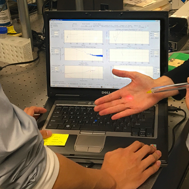
The coagulation monitor consists of a sample probe containing an optical fiber, an optical detector, and a dynamic light signal analyzer. The probe transmits low-coherence light through the optical fiber into the blood. Light scatter signals generated by the circulating red blood cells (in vivo) or blood sample (in vitro) are received by the optical fiber, detected by the optical detector, and analyzed by the dynamic light signal analyzer. The analyzer compares the light scatter signals to a predetermined coagulation value or range and then reports the coagulation status of the blood and/ or clotting time. The system also accommodates multiplexing with other optical assays.
Optical Rheology of Blood during Cardiovascular Surgery, Conference on Lasers and Electro-Optics, OSA Technical Digest, June 2016