
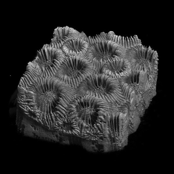
FAU researchers combined X-ray microcomputed tomography with U-Net-based neural networks to analyze the pores and skeletal structures of diseased and healthy stony coral specimens. The non-invasive method proved to be a faster, more accurate way to assess how environmental stressors impact coral skeletons than traditional methods.
The analysis showed how changes in pore structure may compromise skeletal integrity. “Without high-resolution, 3D insights, scientists cannot fully understand how disease, warming oceans and other stressors compromise reef survival,” says Vivian Merk. “By uncovering these hidden changes in porosity, density and skeletal thickness, we can see exactly how diseases like Stony Coral Tissue Loss Disease alter the physical integrity of corals.”
View Related Expert Profiles: Go to Source