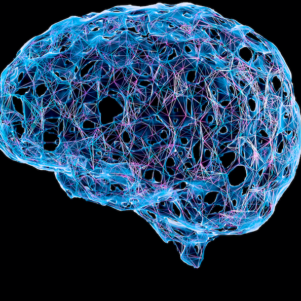
New Interventions May Boost Cognitive Function in HIV Patients
#CognitiveBoost
New Interventions May Boost Cognitive Function in HIV Patients
#CognitiveBoost

Research Terms
Industries
This machine learning system detects or predicts the onset of Alzheimer’s disease by assessing the presence of pathologies associated with the disease in retinal fundus images. Alzheimer’s disease is the most common neurodegenerative disease and is expected to affect 13-16 million people by the year 2050. Alzheimer’s disease causes neural damage that is generally irreversible. Clinical treatment of Alzheimer’s disease focuses on slowing neural degeneration, and early detection is critical for achieving effective treatment. Current detection methods are often not implemented until after the onset of symptoms because they are either invasive, costly, or inaccurate before significant disease progression.
Researchers at the University of Florida have developed a machine-learning algorithm that detects Alzheimer’s disease by analyzing retinal fundus images. After making a disease determination, the algorithm produces a saliency map of the retinal fundus that highlights areas of the image that were critical to making the determination. Unlike current Alzheimer screening methods, retinal images are non-invasive, relatively inexpensive to obtain, and can show Alzheimer’s onset prior to symptoms.
Machine learning algorithm that detects or predicts the onset of Alzheimer’s disease using low-cost, non-invasive retinal images
Retinal fundus images have been studied for their potential to detect Alzheimer’s disease previously, but by-hand measurement and labeling of retinal features is labor intensive and prone to human error. Recent advances in machine learning have used retinal fundus imagery to detect a number of diseases including glaucoma and anemia. This machine-learning algorithm is able to detect or predict the onset of Alzheimer’s disease by examining the retinal vasculature for pathologies associated with the disease.
This non-invasive machine learning software uses fundus retinal images to predict the onset or presence of Parkinson’s disease (PD). Parkinson’s disease is the second most common neurodegenerative disorder, affecting nearly 9 million people, and involves the decay of dopamine production and movement. The gold standard for diagnosing Parkinson’s is the presence of motor symptoms, including tremors, impaired balance, cardiac arrhythmias, and sleep disorders. However, motor symptoms primarily occur in the later stages of the disease when a patient has lost approximately 80% of dopaminergic cells; there is growing interest in exploring non-motor symptoms and biomarkers associated with the disease.
The retina provides a unique opportunity to study neurodegeneration without directly analyzing the brain. In Parkinson’s patients, dopaminergic cells in the substantia nigra, the region of the brain responsible for dopamine and movement, decay and thin the retinal walls and retina microvasculature. The optic nerves are viewable using standard photography or imaging, such as optical coherence tomography (OCT). While OCT imaging provides three-dimensional structures of the eye, fundus eye images are a more clinically relevant diagnosis tool. Retinal fundus images are simply photographs of the back of the eye, including the blood vessels, retina, and optic nerve head, making them highly portable and affordable. It is possible to leverage eye data as a potential means for early Parkinson’s disease diagnosis.
Researchers at the University of Florida have developed machine learning software for identifying the onset or presence of Parkinson's disease by analyzing fundus retinal images. Using a well-trained machine learning algorithm, it identifies vital features in the retinal images, specifically tracking blood vessel patterns, to indicate the presence of Parkinson's in an individual.
Machine learning software analyzes blood vessel patterns in retinal fundus images to diagnose Parkinson’s disease
This non-invasive machine learning software tracks eye blood vessel patterns to diagnose Parkinson’s disease. The software undergoes training using retinal images sourced from two distinct groups: patients previously diagnosed with Parkinson’s and healthy individuals. During the training, the system learns to classify features within the retinal images associated with the two groups. Once training is complete, the system can analyze new retinal images from human subjects. It can detect various retinal features within the retinal image, such as eye blood vessel maps, and predict whether the image indicates an onset or presence of Parkinson’s disease in a patient.
This machine learning algorithm predicts treatment outcome improvements in adults undergoing electrical brain stimulation and enables the delivery of individualized dosing. For example, declines in cognitive function typically present themselves in older adults, who start to naturally experience a decline in functions such as working memory, reasoning, and processing speed. Its prevalence is estimated to be over 11% in adults aged 45 years or older and in almost all adults over the age of 65 years. Transcranial direct current stimulation (tDCS) has been widely used for the last two decades as a therapeutic tool to improve cognitive function, mental health (e.g., depression, anxiety, etc.), and chronic pain in older adults. Its number of applications only continues to grow. However, optimal dosing parameters that underlie positive outcomes, such as electric field intensity or electrode placement, remain evasive. In addition, conventional approaches typically utilize fixed parameters across patients without taking into consideration individual anatomical factors, impacting therapeutic effectiveness.
Researchers at the University of Florida have developed an algorithm that employs machine learning and MRI scans to determine an optimized dosing strategy for the delivery of transcranial direct current stimulation (tDCS), with applications to many forms of electrical brain stimulation (transcranial magnetic stimulation, electroconvulsive therapy, deep brain stimulation, etc.). This information predicts working memory response to tDCS in any individual, enabling precision treatment using individual doses and improving tDCS treatment outcomes.
Machine learning algorithm provides individualized and precise tDCS dosing and predicts treatment outcome improvements from electrical stimulation of the brain
This machine learning algorithm provides precise dosing of electrical stimulation of the brain. By combining finite element computational modeling derived from MRI scans of the brain, identifying tissue types with machine learning, it has the ability to design an optimized dosing strategy for the delivery of electrical current through electrodes on the scalp. This optimized strategy involves subject-specific electrode placement and electric field intensity that can be applied to any new subject so long as a basic MRI scan is acquired.
This software automatically selects the most suitable parameters to develop convolutional neural networks able to denoise medical images. The noise in medical images produced from CT or MRI scans can cover or distort the true image, making accurate diagnosis difficult. The global medical imaging market is projected to exceed $55 billion by 2025. Convolutional neural networks employ deep machine learning that mimics how the brain operates. These networks are useful for analyzing images primarily because of their ability to group images based on similarity and to recognize objects within them. The advantage of using convolutional neural networks to denoise medical images is that they can learn traditionally hand engineered filters without relying on prior work or human input. Available convolutional neural networks for medical image denoising require manual design based on empirical knowledge to eliminate a specific type of noise. However, those denoising algorithms take a long time to create and aren’t efficient due to noise type limitations.
Researchers at the University of Florida have developed a genetic algorithm-based convolutional neural network evolution system in order to improve medical image denoising. This algorithm automatically constructs convolutional neural networks that can effectively denoise a medical image containing complicated and mixed noise types.
Evolutionary deep learning algorithm that automatically constructs convolutional neural networks for medical image denoising
This software is a medical image denoiser development system uses genetic algorithm-based evolution to automate the creation of convolutional neural networks that perform medical image denoising more efficiently than traditional, manually programmed ones. A genetic algorithm is a metaheuristic modeled after natural selection that is most useful for solving complex optimization problems. This system’s genetic algorithm works by exploring the combinations of convolutional neural network parameters and then evolving the most promising of those parameters to create the best image denoiser. The neural networks evolve via selection, crossover, and mutation. This process selects the convolutional neural network most suitable for removing the noise in a medical image. The selected convolutional neural network is able to remove complex noise combinations to improve the accuracy of medical diagnoses based on images from CT and MRI scans. Additionally, this software is applicable in other medical image analyses, such as disease classification and lesion segmentation.
This data-driven framework combines a simulation processor and a denoiser model to transform low-dose, noisy medical scans into high-quality images to reduce patients’ exposure to radiation. While medical imaging is crucial for diagnosing and monitoring diseases, the high radiation doses from conventional scans pose significant health risks to patients and limit the frequency of their use. Currently, the standard practice involves high-dose CT scans that, while effective, expose patients to significant levels of radiation. Annually, approximately 1.6 million people in the U.S. face an increased risk of cancer due to the radiation exposure from CT scans, especially those requiring frequent imaging. However, advancements in low-dose CT technology are transforming the market by reducing these risks while still delivering high-quality images. This growing demand for safer diagnostic tools is driving the CT scan market, which was valued at $8.5 billion in 2023 and is projected to reach $12.8 billion by 2030, as healthcare providers prioritize patient safety alongside diagnostic accuracy.
Researchers at the University of Florida have developed the first medical image restoration model to use phantom and deep learning for real low-dose noise simulation and denoising. This framework involves a machine learning strategy that outperforms state-of-the-art denoising algorithms. It significantly reduces radiation exposure by efficiently denoising low-dose CT scans to produce images of comparable quality to high-dose scans and enhances throughput, allowing physicians to evaluate more patients within the same timeframe.
A data-driven framework that enhances low-dose CT scans by restoring high-quality images, reducing radiation exposure, and speeding up diagnostics for safer, more efficient healthcare
Cyclic Simulation and Denoising is a framework that produces high-quality images from low-dose medical scans, effectively reducing radiation exposure for patients. It combines a simulator, which extracts low-dose noise and tissue features from different image spaces, with a denoiser that effectively reduces the noise while simultaneously restoring tissue details for clearer, more accurate imaging. The cyclic feedback loop between the two components continuously optimizes learning.